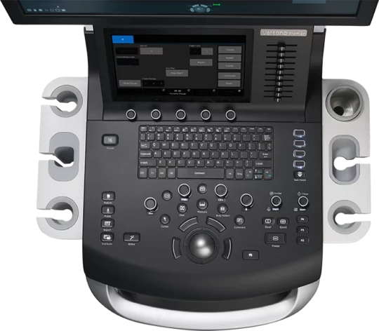
Today’s modern veterinary hospitals incorporate a wide range of imaging technologies that can complement each other and leverage strengths to enhance the diagnostic confidence of busy veterinarians.
Ultrasonography and radiography are pivotal imaging tools in veterinary medicine. Each has distinct advantages that complement each other in diagnosing various conditions. Exploring the differences between these primary imaging technologies helps us understand the benefits of having both in progressive care hospitals.
Radiography excels at providing a global perspective of an animal’s body, particularly bones, organ outlines, lungs, and gas-filled organs like the gastrointestinal tract. Ultrasound enables detailed real-time evaluations of internal organ architecture, free fluid assessments, blood flow characteristics, and directionality, ligament and tendon evaluations, precise needle placements for centesis, aspirates, biopsy or line placements, pericardial space evaluation, and cardiac function, including valve compliance and chamber size assessments, making ultrasound the perfect complement to radiography.
Here are four examples where ultrasound adds additional value when used with digital radiography (DR):
- Confirmation of Abnormal Fluid Accumulation: Ultrasound’s ability to differentiate between fluid and solid tissues makes it invaluable for identifying conditions in the abdomen associated with cancer, bleeding of internal organs causing free fluid, and heart conditions responsible for collapse, like pericardial effusion. It also provides the ability to perform diagnostic and/or therapeutic centesis.
- Differentiating Normal and Abnormal Organs: Ultrasound’s capacity for producing cross-sectional images facilitates precise examination of organ structure, parenchyma, size, blood flow, and vascularity, aiding in the detection of and organ of origin of masses, infections, or infiltrative abnormalities along with the ability to guide diagnostic sampling.
- Heart Disease: Echocardiography, a form of ultrasound, offers comprehensive insights into the heart’s structure and function, valve integrity and chamber size, and the presence of paracardial effusion, which is essential for accurately diagnosing various cardiac conditions.
- Musculoskeletal System: Ultrasound surpasses radiography in evaluating soft tissue structures such as ligaments, tendons, and muscles, making it adept at detecting tears or monitoring healing processes.
- Real-time Imaging: Ultrasound provides real-time imaging, allowing veterinarians to evaluate moving structures, such as dynamic assessments of tendons and ligaments, visual confirmation of therapy delivery, real-time biopsy procedures, blood flow assessments, and comprehensive assessments of cardiac wall motion and valveassessments.6.Portability: Ultrasound machines come in various sizes, from portable handheld devices to larger cart-based systems. The latest portable ultrasound units provide impressive imaging in various clinical settings, including in the field or at remote locations, furtherenhancing ultrasounds’ versatility
While ultrasound offers some unique attributes, digital radiography, and ultrasound work together in the diagnostic toolkit of veterinarians to enhance patient care and improve diagnostic confidence.

