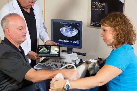
Frequently asked questions regarding ultrasound, Q&A
Ultrasound is the second most used imaging technology in veterinary hospitals. Its versatility, combined with real-time imaging of soft tissue structures, gives Veterinarians great insight into an animal’s clinical well-being. This powerful imaging tool has a purpose in every veterinary hospital. Let’s learn more about this exciting modality and its purpose.
How does ultrasound work?
Ultrasound imaging, also known as sonography, works on the principle of sound waves. Here’s a simplified breakdown of how it operates:
- Generating Sound Waves: The process begins with a transducer containingpiezoelectric crystals. When an electric current passes through these crystals, they vibraterapidly, generating sound waves at frequencies higher than the human ear candetect(typically in the range of 1 to 22 megahertz).
- Sound Wave Transmission: Good sound wave transmission happens when a couplingagent like gel is placed between the transducer and the skin.Shaved animals provide thebest image quality.These sound waves travel from the transducer into the body. Theypenetrate through the skin and other tissues, encountering different tissue types along theway, such as muscle, fluid, or organs.
- Reflections and Echoes: When the sound waves encounter a boundary betweentissuesor a structure that reflects sound differently (like bones), some waves are reflected backtowards the transducer. The amount of reflection depends on the density and compositionof the tissue.
- Receiving Echoes: The transducer’s crystals detect the returning sound waves, which now act as receivers. They convert the mechanical energy of the returning waves backinto electrical signals.
- Image Formation: The electrical signals containing information about the echoes aresent to the beamformer, which analyzes them. The processed data is used to generate agrayscale image representing the body’s internal structures. Brighter areas on the imagetypically represent stronger echoes (such as from dense tissues), while darker areasrepresent weaker ones (suchas fluid-filled regions).The other component of theprocessed data measures the time it takes for the echoes to return from the body; thesystem can determine the distance to the tissue boundary and its position on the display.
- Image Display: The resulting image is displayed on the ultrasound machine’s monitor inreal time. Healthcare professionals can interpret these images to diagnose conditions,monitor disease states and fetal development, guide medical procedures, and more.
Why are there different types of transducers?
Different types of ultrasound transducers exist because various medical imaging needs require different characteristics and capabilities. Here’s why;
- Applications (shape and size): Transducers are designed for specific medical applications. For example, linear transducers are often used for near-field imaging like musculoskeletal, thyroid, and abdominal imaging in cats and smaller dogs. In contrast, curvilinear or phased array transducers are preferred for abdominal and pelvic applications, and sector transducers are exclusively used for cardiac imaging. Endocavitary transducers are designed for imaging within body cavities like the rectum or vagina. Trans Esophageal transducers are used to evaluate the heart from inside the esophagus
- Frequency Range: Transducers come in different frequency ranges, typically 1 to 22megahertz (MHz). Higher frequencies provide better resolution for superficial structures like small parts like thyroids, blood vessels, and tendons, while lower frequencies penetrate deeper into the body to image abdominal organs and hearts in larger patients.
- Special Features: Some transducers include special features like matrix 3D/4D imaging capabilities, remote operation of system controls via probe buttons, form factor designs that improve access for tight spaces such as surgery, transducers with increased field of view to observe more anatomy, and needle guidance brackets for biopsies, therapy placement or line placement. These additional features cater to specific diagnostic needs and enhance the utility of the transducer in clinical practice.
- Patient Comfort and Accessibility: Transducers may vary in design to enhance patient comfort and accessibility during imaging. For example, transducers designed for smaller animal imaging will have smaller footprints and smoother edges to minimize discomfort for petite patients.
- Technology Advancements: With technological advancements, newer transducers may incorporate features such as matrix array technology and single crystal technology, which allows for better beamforming and improved image quality, or wireless connectivity for increased flexibility and ease of use.
Overall, the diversity in ultrasound transducers allows healthcare providers to tailor imaging procedures to specific patient needs, clinical scenarios, and diagnostic requirements, ultimately facilitating more accurate diagnoses and better patient care.
What can ultrasound be used for in veterinary hospitals?
Animals may need an ultrasound for various reasons. Here are some everyday situations.
- Trauma: Assessment of internal bleeding (free fluid), organ damage, soft tissue injuries(hematomas, tissue tears), and fracture evaluations. While not a primary imaging modality for evaluating fractures, ultrasound can provide additional information about soft tissue involvement around fractures like hematomas. Pneumothorax in chest trauma, organ perfusion specifically infracts, and necrosis in traumatized kidneys. Guidance for procedures like draining fluid collections like abscesses and hematomas or biopsy of suspicious tissues, needle, and catheter placement, and guidance ensuring precisetargeting and minimizing complications.
- Abdominal Imaging: Ultrasound is commonly employed to assess abdominal organs such as the liver, spleen, kidneys, bladder, adrenals, and gastrointestinal tract. It helps diagnose conditions like tumors, cysts, obstructions, and inflammation.
- Cardiac Evaluation: Echocardiography, a specialized form of ultrasound, assesses animal heart health. It evaluates cardiac structure, function, and blood flow, aiding in the diagnosis of congenital defects, valvular disease, cardiomyopathies, pericardial effusion, and other cardiac conditions.
- Postoperative Monitoring: Ultrasound allows veterinarians to monitor the surgical site and surrounding tissues for signs of complications such as hematoma formation, seroma formation (accumulation of fluid), or abscess formation. It helps ensure proper healing and timely intervention if complications arise.
- Guidance for Drain Placement: If drainage is necessary post-surgery to remove fluid or prevent fluid accumulation, ultrasound can guide the placement of drainage catheters or tubes, ensuring accurate positioning and effective drainage.
- Localization of Foreign Bodies: Ultrasound can effectively locate foreign bodies within the GI tract or other anatomical structures. The characteristic appearance of foreign bodies on ultrasound varies depending on their composition and location, but they often appear as hyperechoic (bright) structures with distal acoustic shadowing.7.Ocular Evaluation: In veterinary ophthalmology, ultrasound is used to evaluate ocular structures, particularly in cases of opaque media or when direct visualization is challenging. It helps diagnose intraocular tumors, retinal detachment, and lens luxation.
Thoracic Imaging: Ultrasound can assess thoracic organs such as the lungs, pleura, and mediastinum. It aids in diagnosing conditions like pleural effusion, pneumothorax, pulmonary masses, and mediastinal tumors.

