
Ultrasound has become an indispensable tool in veterinary medicine. It is a non-invasive, real-time diagnostic option that provides detailed insights into animals’ internal structures. Its vast applications encompass everything from routine health checks to emergency diagnostics. Here, we delve into some common indications for using ultrasound in veterinary general practice.
Abdominal Imaging
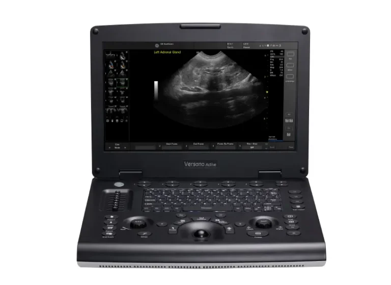
One of the most frequent uses of ultrasound in veterinary practice is abdominal imaging. The liver, kidneys, bladder, gastrointestinal tract, spleen, adrenal glands, abdominal lymph nodes, and pancreas can all be examined using ultrasound.
- Liver Evaluation: Ultrasound can detect abnormalities such as an increase or decrease in echogenicity, nodules, tumors, cysts, or abscesses. It helps assess liver margins, texture, and the presence of lesions, offering a clearer picture than conventional radiographs. It can also guide cytologic sampling.
- Kidney Assessment: Conditions like hydronephrosis, cysts, mineralization, or kidney tumors are often diagnosed with ultrasound. This modality allows for a detailed examination of kidney structure, supporting in the diagnosis of chronic kidney disease or acute kidney injury.
- Bladder and Urethra Examination: Ultrasound is crucial for detecting stones, debris, tumors, or inflammation in the bladder and urethra. It also helps in identifying obstructions or anomalies that might cause urinary issues.
- Gastrointestinal Tract: The stomach, intestines, and associated structures can be evaluated for masses, foreign bodies, changes in wall layers, or motility issues. Ultrasound helps in diagnosing conditions such as gastrointestinal tumors, obstruction, and inflammatory diseases.
- Spleen Evaluation: An enlarged spleen or splenomegaly, as well as nodules, masses, or infarctions, can be identified using ultrasound, providing vital information for diagnosing systemic diseases or trauma, as well as guide cytologic sampling.
- Adrenal Glands: Evaluating adrenal size and the presence or absence of nodules or masses may assist in the diagnosis of hyperadrenocorticism, hypoadrenocorticism, or cranial retroperitoneal masses (e.g., adenocarcinoma, or neuroendocrine tumors). It may also evaluate for local infiltration of the caudal vena cava, kidneys, or sublumbarmusculature.
- Pancreas Examination: Diagnosing pancreatitis- acute or chronic is significantly aided by ultrasound, which is sensitive to inflammatory changes, changes to the pancreatic duct, pancreatic parenchyma, and duodenal papilla.
- Lymph Nodes: Enlarged lymph nodes, often a sign of infection or cancer, can be examined using ultrasound to assess their size, structure, and any associated abnormalities and guide cytologic sampling.
Cardiac Imaging (Echocardiography)
Echocardiography, or cardiac ultrasound, is another vital application in veterinary practice, particularly for diagnosing heart diseases.
- Heart Disease: Ultrasound assesses heart size, wall thickness, and valve function, making it invaluable for diagnosing and monitoring conditions like cardiomyopathy and valvular diseases.
- Pericardial Effusion: This condition, characterized by fluid accumulation around the heart, is readily identified through ultrasound, allowing for timely intervention.
- Congenital Heart Defects: Structural abnormalities present from birth can be diagnosed early with ultrasound, guiding treatment plans, and surgical interventions.
Reproductive Imaging
Reproductive health is another area where ultrasound shines, providing essential information for breeding management and reproductive health monitoring.
- Pregnancy Diagnosis and Monitoring: Ultrasound confirms pregnancy, monitors fetal development, and helps detect any complications early in the gestation period. It is also used to estimate fetal viability and staging for c-section when indicated. Remember to obtain radiographs at >45 days of pregnancy for accurate fetal count.
- Uterine and Ovarian Health: Conditions like pyometra, ovarian remnant, or tumors are diagnosed through ultrasound, enabling prompt and appropriate treatment.
- Testicular and Prostate Evaluation: Ultrasound helps in identifying tumors, cysts, or inflammation in the testicles and prostate, aiding in the diagnosis of reproductive and urinary tract disorders.
Thoracic Imaging
While radiographs are typically the first choice for thoracic imaging, ultrasound provides valuable complementary information, especially for soft tissue evaluation.
- Pleural Effusion: Ultrasound is effective in detecting and characterizing fluid accumulation in the pleural space, which can result from infections, heart failure, or trauma. It is also useful in guiding diagnostic and/or therapeutic thoracocentesis.
- Lung Masses or Lesions: Ultrasound helps in identifying, assessing, and sampling tumors or other abnormalities within the lungs, although air-filled structures limit its penetration.
- Diaphragmatic Hernia: This condition, in which abdominal organs protrude into the thoracic cavity through a defect in the diaphragm, can be diagnosed with ultrasound.
Soft Tissue Imaging
Ultrasound is extensively used for imaging soft tissues, providing a detailed assessment of structures beneath the skin.
- Skin and Subcutaneous Structures: Masses, cysts, or abscesses in the skin or just below it can be evaluated using ultrasound, guiding further diagnostic procedures, sampling, or treatment.
Emergency Situations
In emergency veterinary medicine, ultrasound is critical in rapid assessment and diagnosis.
- Trauma Assessment: Ultrasound helps in quickly evaluating internal bleeding, organ damage, and other traumatic injuries, guiding emergency interventions.
- Foreign Body Detection: Ingested or embedded foreign objects can be located and identified using ultrasound, which is complemented by radiographs for a global perspective, aiding in their safe removal.
Guidance for Procedures
Ultrasound also serves as a guide for various diagnostic and therapeutic procedures.
- Fine Needle Aspiration/Biopsies: Ultrasound guidance ensures accurate needle placement for tissue sampling, improving diagnostic yield and safety.
- Fluid Drainage: Draining abscesses or effusions is facilitated by ultrasound, ensuring precision and reducing the risk of complications.
Conclusion
In veterinary general practice, ultrasound is a versatile and invaluable diagnostic tool. Its ability to provide real-time, detailed images of internal structures makes it essential for diagnosing a wide range of conditions, from routine health checks to emergency diagnostics. By enhancing the ability to characterize and assess various organs and tissues, ultrasound significantly improves the quality of care provided to animal patients.