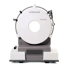
Computed Tomography (CT) has revolutionized diagnostic imaging in veterinary medicine, offering 3-dimensional imaging and multiphasic contrast study ability compared to traditional 2-dimensional modalities like X-ray and ultrasound. Particularly in small animal practice, CT provides an additional tool for veterinarians, enabling them to image complex cases in the clinic with greater precision and confidence. As veterinary medicine continues to advance, the adoption of CT technology has grown, reflecting its substantial benefits in effectively managing animal health. This analysis explores the diagnostic capabilities of CT, supported by specific case studies and practical applications in general practice, providing a comprehensive view for veterinary professionals.
Diagnostic Capabilities of CTCT scanning offers a myriad of diagnostic advantages, particularly significant in the context of small animal health. Unlike standard radiography, which may be limited due to the superimposition of structures, CT images allow for a three-dimensional evaluation and reconstruction of the animal’s anatomy. This capability is crucial for accurately assessing complex anatomical regions such as the skull, spine, and joints.
- High-Resolution Imaging: CT scans produce high-resolution images that can detect minuted differences in tissue density. These images are essential for identifying subtle fractures, small tumors, and early signs of disease that may not be visible on traditional X-rays.
- Speed and Precision: Modern multirow detector CT (MDCT) scanners can complete a full body scan in under a minute. This speed minimizes the time animals need to be under anesthesia, reducing risk and discomfort. The quick turnaround time is vital in emergency situations where a rapid diagnosis is essential to providing high-level patient care.
- Enhanced Contrast: Using contrast agents in MDCT imaging can highlight vascular structures and differentiate between tissue types. This is particularly useful in oncology cases, where distinguishing between tumor tissue and normal tissue is critical for accurate diagnosis and treatment planning.
- Comprehensive Neurologic Evaluation: MDCT is highly effective for diagnosing neurologic conditions. It can be used to localize and confirm spinal compression and evaluate for hemorrhage, tumors, and other anomalies within the skull and spinal cord, which are often difficult to evaluate fully with 2-dimensional diagnostic tools. The advanced diagnostic capabilities of CT significantly enhance veterinarians’ ability to diagnose and treat a wide range of conditions more effectively. The following sections will delve deeper into real-world applications through detailed case studies, illustrating the practical uses and benefits of CT in a small animal veterinary practice.
Case Studies
1. Intracranial Tumor in a Small Breed Dog
In a compelling case at a veterinary hospital, a 6-year-old Poodle presented with a history of seizures and behavioral changes. Traditional radiography and ultrasound excluded thoracic or abdominal causes, prompting the veterinary team to perform a CT scan. The CT images (pre and post-intravenous contrast) revealed a small, well-defined mass in the left cerebral hemisphere. The high-resolution CT scan allowed for precise localization and assessment of the tumor’s size and its effect on surrounding tissues, facilitating targeted surgical planning. The dog showed significant improvement post-surgery, underscoring the CT’s crucial role in managing complex neurological disorders.
2. Complex Abdominal Issues in a Cat
A 4-year-old Siamese cat was brought in with vague symptoms, including lethargy and intermittent vomiting. An abdominal ultrasound demonstrated irregularities in organ texture. Subsequent CT scanning provided a detailed view of the abdominal organs, revealing multiple small nodules throughout the liver and abnormal enlargement of the pancreas. These findings led to a diagnosis of metastatic pancreatic cancer, guiding the veterinary team toward appropriate palliative care and informing the owners about the prognosis and treatment options.
3. Nasal Case in a Dog
A Golden Retriever exhibited chronic nasal discharge and episodic nosebleeds, symptoms commonly associated with differential diagnoses such as fungal infections, foreign bodies, or tumors. Nasal swabs and radiographs did not yield conclusive results. A CT scan of the nasal cavities was performed, which identified a foreign object lodged deep within the nasal passage, surrounded by minor tissue inflammation. The object, a small piece of wood, was surgically removed, and the dog recovered fully. This case illustrates how CT can be indispensable in diagnosing cases where conventional methods have not yielded a diagnosis.
4. Thoracic Case in a Cat
In a case involving a 9-year-old Domestic short-haired cat exhibiting respiratory distress, a thoracic radiograph demonstrated a mass in the thoracic cavity with an indeterminate pleural, pulmonary, or mediastinal location. A CT scan provided a clear and detailed three-dimensional view, confirming the presence of a large, invasive thymoma compressing the lung tissue. This precise imaging allowed for a strategic surgical approach, which resulted in the successful removal of the tumor and significant improvement in the cat’s breathing and overall quality of life.
5. Metastatic Evaluation in a Dog
A 10-year-old Boxer with a history of melanoma was evaluated for potential metastasis. Given the likelihood of metastasis, a CT scan was performed to check for metastatic lesions throughout the body. The CT revealed several small lesions in the lungs and liver, consistent with metastasis. This comprehensive evaluation was crucial for directing tissue sampling for confirmation of metastasis and adjusting the ongoing treatment plan, emphasizing the role of CTin monitoring and managing cancer progression in veterinary patients. These cases highlight the diverse applications of CT imaging in diagnosing and managing complex medical conditions in small animals, showcasing its indispensable value in general veterinary practice. Next, we will discuss the practical use cases and cost-benefit analysis of CT technology in general practice.
Practical Use Cases in General Practice
When to Use CT Imaging
CT imaging is particularly beneficial in scenarios where traditional diagnostic methods may be limited. Here are vital situations where CT can be advantageous to a high level of patient care and clinical practice
- Complex Diagnoses: CT should be considered when diseases involve complex anatomical regions like the brain, nasal cavities, middle and inner ear, spinal column, or joints, where detailed cross-sectional imaging is necessary.
- Trauma Cases: CT can quickly provide a comprehensive overview of internal injuries for animals involved in trauma, aiding in rapid and effective treatment planning
- Cancer Evaluation: CT is invaluable for cancer staging. It helps to identify the extent of the disease and the involvement of surrounding tissues, which is crucial for determining appropriate treatment protocols and prognosis for clients.
- Chronic Diseases: In cases of chronic or recurring symptoms where other modalities have not provided conclusive information, CT can offer new information and help refine treatment strategies.
Guidelines for General Practitioners
- Training and Understanding: Ensure staff are adequately trained and understand when CT is the most appropriate imaging choice.
- Referral Relationships: Develop relationships with referral centers with CT capabilities if direct access to a CT scanner is unavailable.
- Cost Management: Discuss the cost implications with pet owners, providing clear information on the benefits and potential diagnostic outcomes.
Cost vs. Benefit Analysis
Assessing the Costs
The investment in CT technology can be significant, with the initial equipment cost and the need for ongoing maintenance and training. However, recruiting and retaining cases, providing enhanced clinical care, and increasing caseload and revenue can offset these costs over time.
Benefits of CT Scanning
- Improved Diagnostic Accuracy: CT scanning provides clear, precise images that can lead to more accurate diagnoses, reducing the need for repeat testing and unnecessary treatments.
- Enhanced Treatment Outcomes: With better diagnostic data, treatment plans can be more targeted and effective, improving patient outcomes.
- Client Satisfaction: Offering advanced diagnostic options can improve client trust and satisfaction, as clients appreciate the thorough care provided to their pets.
Return on Investment
While the upfront costs are high, the return on investment can be substantial in financial terms and the enhanced level of care provided. Practices that invest in CT technology may see an increase in referrals and a higher standard of diagnostic services, attracting more clients and enhancing the practice’s reputation.
Conclusion
CT technology represents a significant advancement in veterinary diagnostic imaging. Its capabilities to provide detailed, accurate images make it an indispensable tool in diagnosing and treating a wide range of conditions in small animals. While there are investments, enhanced diagnostic accuracy, better patient outcomes, and improved client satisfaction make CT an invaluable addition to veterinary practices aimed at providing the highest standard of care. As the technology becomes more accessible, more general practices will likely consider integrating CT scanning into their array of diagnostic tools, further enhancing the quality of veterinary care.

